Ultrasound for shoulder pain
Shoulder pain that persists during exercise, lifting or sleeping requires more than just guesswork. We use ultrasound to identify exactly which parts of the shoulder are causing the problems, so that we can start the right treatment plan as quickly as possible to help you get back on your feet. The shoulder is a complex joint where conditions often resemble one another and can produce similar symptoms.
When should an ultrasound scan be carried out for shoulder pain?
Shoulder problems can originate from various structures. The rotator cuff, the bursa, the long biceps tendon or the AC joint can all cause pain. The difficulty is that, in practice, these symptoms often resemble one another. Pain on the outside of the shoulder, difficulty with overhead movements and night-time pain are common to several different conditions.
It is precisely in these cases that an ultrasound scan is useful. An ultrasound scan allows us to examine tendons, muscles, bursae and other structures around the shoulder quickly and precisely. This is particularly useful if the symptoms have been present for some time, if the pain persists during everyday activities, or if there is uncertainty as to whether the problem is, for example, tendon irritation, bursitis or even a tear.
Following a fall or a sudden loss of strength, an ultrasound scan can also help to assess whether there is actually any damage to a tendon. Think of situations where you suddenly find it difficult to lift something, or where a particular movement causes immediate, sharp pain. In such cases, you don’t want general advice, but clear information. In some cases, referral to hospital via your GP may be an option.
What can we see on an ultrasound scan of the shoulder?
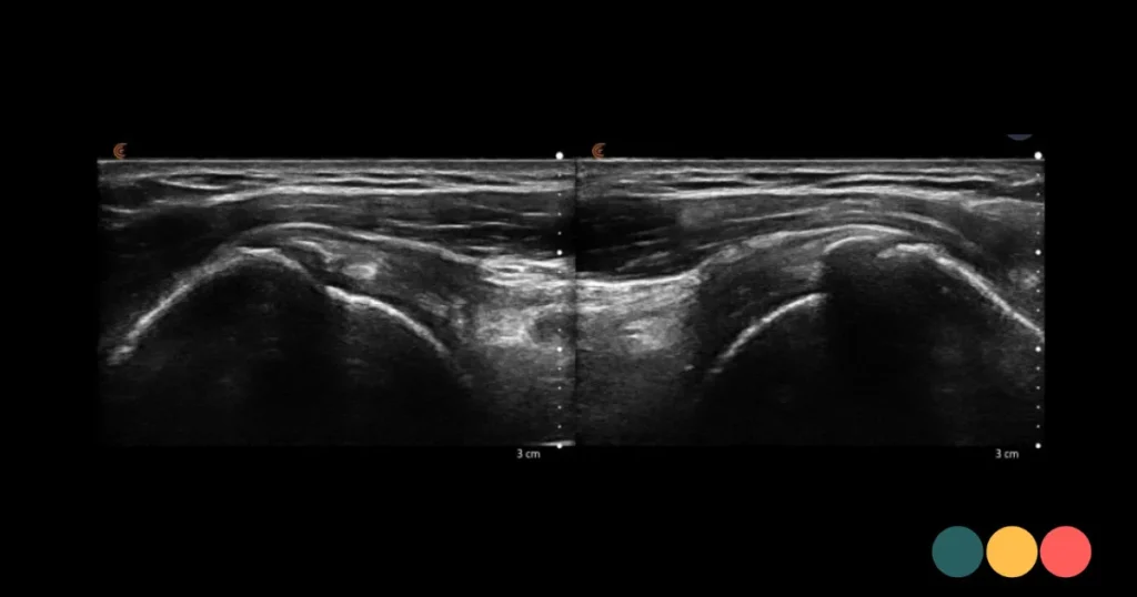
An ultrasound scan is particularly suitable for visualising superficial soft tissues. In the shoulder, we often examine the tendons of the supraspinatus, infraspinatus and subscapularis muscles, as well as the biceps tendon. A thickened or inflamed bursa is also usually clearly visible.
This makes ultrasound particularly useful for conditions such as tendinopathy, calcification, bursitis or (partial) tendon tears. Sometimes we also observe fluid, irritation or changes in tendon structure that are more consistent with long-term overuse. This information helps us to better assess the severity of the condition and the patient’s capacity for physical activity.
What an ultrasound scan does not show as clearly are deeper structures such as cartilage, the labrum, or problems originating more from within the joint itself. So there is an important nuance to bear in mind right away. A normal ultrasound scan does not automatically mean that there is nothing wrong. It means that the soft tissues examined do not show any clear abnormalities. Research and technology are constantly improving, and, with due caution, we are also able to reach a clear diagnosis regarding these structures with increasing certainty.
An ultrasound scan is never carried out in isolation from a physical examination. The findings must be consistent with how you feel, what we are testing for, and how the symptoms developed.
Why rapid diagnosis of shoulder pain makes all the difference
People often carry on for too long with a shoulder that keeps flaring up. First a bit of rest, then a fresh attempt, then the pain returns. This is a pattern we often see in athletes, people in physically demanding jobs and those whose work involves a lot of activity above shoulder height. Without a clear diagnosis, recovery can become a bit of a mess in some cases.
Bursitis, for example, requires a different treatment plan to a degenerative tendon condition or a tear in the rotator cuff. Our expectations regarding a return to work, sport and a good night’s sleep also change once we have a better understanding of what is going on.
That clarity often has a significant impact on a mental level as well. If you understand why a particular movement causes pain and what is still safe to do, you’ll move with greater confidence. That’s important, because uncertainty This often leads to people either avoiding a particular movement altogether or, conversely, putting too much strain on themselves. We often see an ‘all or nothing’ mentality. .
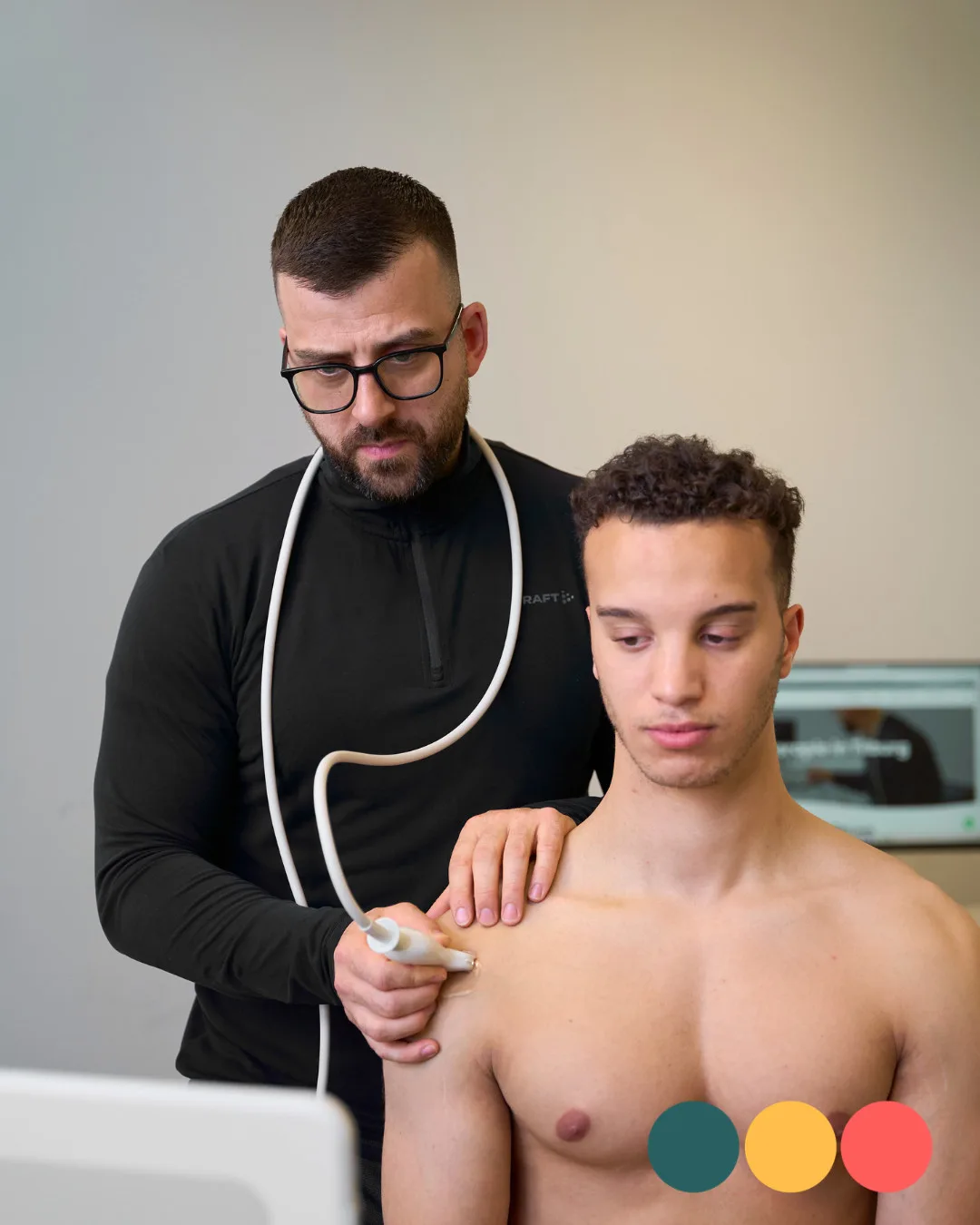
How is an ultrasound scan of the shoulder carried out?
A shoulder scan is straightforward and usually doesn’t take long. You don’t need to do anything special to prepare for it. During the examination, we examine the shoulder in various positions and often whilst it is moving. The latter is a major advantage of ultrasound. We can see not only what it looks like at rest, but also what happens during active movement.
This dynamic assessment is very important in cases of shoulder complaints. Some irritations only become apparent when the tendon moves beneath the acromion or when a bursa becomes pinched during lifting. This makes an ultrasound scan both practical and clinically valuable.
We often compare the affected side with the other shoulder as well, especially if the findings aren’t immediately clear. And once again, not every abnormality seen on an ultrasound scan is automatically the cause of your pain. In adults in particular, we see changes in the tendons that are normal and do not necessarily cause any immediate symptoms. That is why interpreting all the information remains more important than just the image itself.
What shoulder problems do we often see on an ultrasound scan?
One of the most common findings is tendinopathy of the supraspinatus tendon. This is a strain-related condition affecting the tendon, often causing pain when lifting, reaching and sleeping on that side. We also regularly see bursitis, which can be particularly painful during overhead movements.
In addition, calcium deposits can form in tendons. These can be extremely painful, but not every case of calcification explains all the symptoms. The size, location and condition of the deposits help determine how significant such a finding is. This is precisely why an ultrasound scan carried out by a physiotherapist or specialist with expertise in the musculoskeletal system offers added value. Experience is therefore important.
Partial or complete tears of the rotator cuff may also be visible. In athletes, this sometimes occurs acutely following trauma, whilst in older patients it is more often the result of wear and tear or long-term overuse, and is frequently preceded by a fall. Treatment therefore varies. Age, activity level and loss of function are all factors in determining whether a referral is appropriate.
An ultrasound scan for a shoulder complaint is not an end in itself
A good ultrasound scan is not an end in itself, but it provides information that will hopefully have a positive impact on your recovery process. The real question is not just what can be seen, but what you need to be able to move, work or exercise normally again. This is where the process of translating the images into a plan begins.
If the ultrasound scan indicates an overloaded tendon, recovery often involves temporarily adjusting the load, improving shoulder control and gradually building up strength. In the case of bursitis, the initial focus may be on pain relief and restoring normal range of motion, followed by strength and load-bearing capacity. In the event of a tear or obvious rupture, further medical assessment may sometimes be required.
What is the difference between an MRI and an X-ray?
People often ask whether an MRI isn’t a better option. That depends on the specific issue. For tendons, bursae and dynamic assessments, ultrasound is often quick, effective and can be used straight away. What’s more, during the scan you can mimic the painful movement and see what’s happening in real time.
An MRI provides a more detailed view of deeper structures and more complex joint problems. Examples include a labrum injury or cases where previous investigations have failed to explain persistent symptoms. An X-ray, on the other hand, focuses more on bone structures, osteoarthritis or alignment abnormalities, and less on tendons and bursae.
The best option therefore depends on the symptoms. Not every shoulder requires an X-ray or MRI straight away. Particularly in the case of common tendon and bursa problems, ultrasound is often a logical first step and a cost-effective option.
When should you get in touch to book an ultrasound scan?
If your shoulder pain persists for longer than a few weeks, disrupts your sleep, causes a noticeable loss of strength, or makes it almost impossible to move your arm above your head, further investigation may be useful. This also applies if the pain keeps returning despite rest or exercises you have already tried.
In the event of an acute injury accompanied by an audible snap or click, significant weakness or an inability to lift the arm properly, you want clarity as soon as possible. In such cases, it is important to know whether there is any significant tendon damage and what the next step should be.
Putting up with an unclear shoulder problem often takes longer than having it properly assessed. Especially if you want to get back to sport, work or simply sleep without pain, a quick diagnosis will help you get back on track.
Related complaints

Biceps tendon rupture
Tearing of the biceps tendon usually happens while the biceps is applying too much sudden force...
Cuff rupture in the shoulder
Do you have pain, loss of strength or difficulty lifting your arm? Then the...
Deltoid tendinopathy
A deltoid tendinopathy occurs due to reduction in quality of tendon tissue.