Calf injuries: from whiplash to tendon injury this determines how soon you will be back on the field
Injuries to the calf are common during explosive sports such as football, running and tennis. The most common calf injury is a muscle tear, also known as whiplash. This often causes acute pain at the back of the calf. Very recognisable is the idea of having been tapped by an opponent when often no one is around. Whiplash may seem like a simple muscle injury, but recovery depends greatly on the exact location and type of tissue damaged. Where previously it was thought that 3 weeks of rest was enough, we now know better. With a more focused diagnosis and a thoughtful treatment plan, you will return to the field safe and strong, without the risk of another injury.

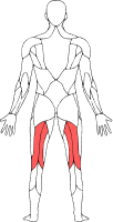
General anatomy of the calf
The calf region is formed by a group of muscles collectively known as the triceps surae (tri- = three, ceps = heads, surae = calf) are called. It consists of:
- Gastrocnemius: the superficial calf muscle with two heads (medial and lateral) that spring from the femur. The medial head is often involved in whiplash injuries.
- Soleus: a deeper calf muscle that lies below the gastrocnemius and together with it forms the Achilles tendon.
- Plantaris: a small, thin muscle with a long tendon that is absent in some people. May also be affected by whiplash.
The muscle bellies of the gastrocnemius and soleus merge into aponeuroses (flat tendon-like structures), which eventually come together and form the Achilles tendon forms. Knowledge of this anatomy is important to properly classify and treat injuries.
Besides the muscle structures, there are many other tissues in the calf region that are relevant in the occurrence or recognition of injuries. These include the fadpad, bursas, nerves, blood vessels and other fascial structures.
Thus, fat bodies such as the retrocalcaneal fat pad provide shock absorption in the area between skin, tendons and bone. Bursas play a role in smooth movement can become irritated with repeated movement or pressure. Nerves such as the sural nerve can cause symptoms due to local swelling or scar tissue. Other connections between muscle groups, such as the deep fascial compartment of the lower leg, can also cause complaints and cause local pain.

What actually is a calf lash
In this blog, we focus specifically on the gastrocnemius, as this muscle head is most often involved in whiplash. The gastrocnemius is bi-articular, meaning that this muscle runs over both the knee joint and the ankle joint. This makes this muscle especially vulnerable during movements involving simultaneous flexion of the ankle and extension of the knee - a typical situation when sprinting or having to stop abruptly. Because of this function, the load on the gastrocnemius is high, increasing the risk of injury. In particular, the medial, or inner, head. The medial head is more often affected because of the anatomical and functional differences between the medial and lateral heads. The medial head is slightly longer, more powerful and has a larger pennation angle, which means it experiences more tensile strain under eccentric loads (such as sudden braking or overstretching). In addition, the transition between muscle and aponeurosis is more sharply defined in the medial head. This makes the medial head more susceptible to rupture during explosive movements.
A whiplash refers to acute pain and functional impairment in the calf region caused by a tear, in most cases of the medial head of the gastrocnemius. However, the plantaris or soleus may also be involved. The term whiplash refers to the feeling often described by athletes: as if someone struck the calf with a whip. The well-known tap, in other words.
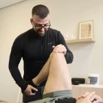
Often but not always, we see marked swelling, sometimes a haematoma(haematoma) or blue discolouration. It is also often painful with dorsal flexion of the ankle in combination with knee extension. Palpation usually shows pressure sensitivity of the calf, sometimes with a palpable dent in the muscle belly.

From complaint to clarity
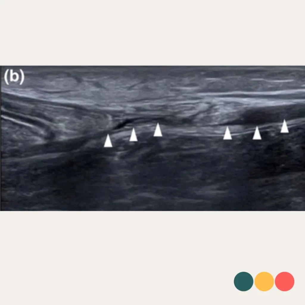
Ultrasound is a quick and accessible way to properly map this injury in the diagnosis and differentiation of calf injuries, and particularly in injuries of the medial gastrocnemius. Thanks to its ability to assess dynamically - for example, during dorsiflexion and plantarflexion - not only the location of the injury, but also the degree of muscle function can be revealed. This makes it very valuable in determining the severity of an injury as well as predicting recovery time.
Injuries of the gastrocnemius vary significantly depending on the tissue type involved. By looking not only at the size of the injury, but also at the exact location within the muscle-tendon continuum (muscle fibre, transition zone, aponeurosis or free aponeurosis), A proper diagnosis is crucial to differentiate between these areas. Below is an overview of the four most common injury types of the medial gastrocnemius, each with its own pattern and prognosis.
Type 1 - Myoaponeurotic lesion without aponeurotic defect
- Localisation: transition zone muscle fibre/perimysium to aponeurosis
- Aponeurosis: intact
- Haematoma: none
- Movement: synchronous
- Prognosis: favourable, short recovery time (usually <2 weeks)
- Indication of mild rupture without interruption of load-bearing structures.
Type 2 - Aponeurotic lesion of the gastrocnemius aponeurosis (GA)
2A less than 50% of gastrocnemius aponeurosis affected(transverse shot)
- Haematoma: mild to moderate
- Movement: synchronous
- Prognosis: average recovery (2-4 weeks)
- Recovery is often possible conservatively with progressive loading.
2B more than 50% of the gastronemicus aponeurosis affected(transverse uptake)
- Haematoma: often moderate to severe
- Movement: asynchronous
- Prognosis: longer recovery time (4-6 weeks or longer)
- Monitoring of muscle activity and ultrasound follow-up recommended.
Type 3 - Injury of the free gastrocnemius aponeurosis
Tendon injury closer to attachment on Achilles tendon
- Haematoma: none or mild
- Movement: mostly synchronous
- Prognosis: tendinogenic healing → often longer recovery (average 5-7 weeks)
- Usually requires a longer build-up in training and load
Type 4 - Mixed injury
- Both aponeurosis and the free tendon are affected
- Haematoma: almost always present
- Movement: asynchronous
- Prognosis: worst outcome, average recovery + 6 weeks
- It can take months for full return to sport, especially in athletes with high load factors.
Type 1: rapid return to sport (10-14 days on average)
Type 2A: 21 days on average
Type 2B: 35-40 days on average
Type 3: 5-7 weeks on average
Type 4: > 6-8 weeks
The study shows that a single snapshot with ultrasound can tell a surprising amount about the severity of a calf injury. For example, more severe muscle injuries (such as type 2B and type 4) showed that the gastrocnemius and soleus no longer cooperate synchronously during movement. Also, in these types, a haematoma (bleeding) was often seen between the muscle layers - a sign of substantial tissue damage. Based on these characteristics and clinical experience, clear differences in recovery time could be identified: from an average of two weeks in type 1 to over six to eight weeks in type 4.
Clarity from the start, less relapse, more progress
This classification gives physiotherapists and sports physicians a concrete tool to set the right expectations and better manage the rehabilitation process. Not every muscle injury is the same and this requires a tailored approach. Understanding the severity of the injury at an early stage creates more peace, clarity and confidence in the recovery process and reduces the likelihood of relapse.
Source: Pedret C, Balius R, Blasi M, et al. Ultrasound classification of medial gastrocnemious injuries. Scand J Med Sci Sports. 2020;00:1-10. https://onlinelibrary.wiley.com/doi/10.1111/sms.13812
Related complaints

Achilles tendon rupture
A complete rupture of the Achilles tendon often occurs during explosive movements where there is a sudden application of a lot of force,...
Achilles tendon problems in runners
Achilles tendon complaints are a common problem in people who are active in sports ...
Hamstring complaints in runners
Hamstring complaints are common among runners. Pain and loss of strength are common symptoms in hamstring injuries.