Ultrasound in tendonitis: What do you see and what can you do with it?
Tendon pain is common and can be persistent. Think of pain around the Achilles tendon, shoulder, knee or elbow. These complaints can significantly affect daily functioning, especially with sports or repetitive movements. But how do you know exactly what is going on? Ultrasound can help.
What are tendon complaints?
Tendon injuries usually occur due to overuse, degeneration or (micro)trauma. The tendon becomes irritated or damaged, leading to pain, stiffness and sometimes swelling. Common tendon complaints are:
- Achilles tendinopathy
- Shoulder (rotator cuff) tendinopathy or calcification
- Jumpers knee (patellar tendonitis)
- Tennis elbow (extensor tendons)
Risk factors
Factors that increase the likelihood of tendon problems:
- Prolonged overexertion or repetitive motion
- Poor technique or posture
- Reduced muscle strength or mobility
- Aging tendon tissue
- Obesity and smoking
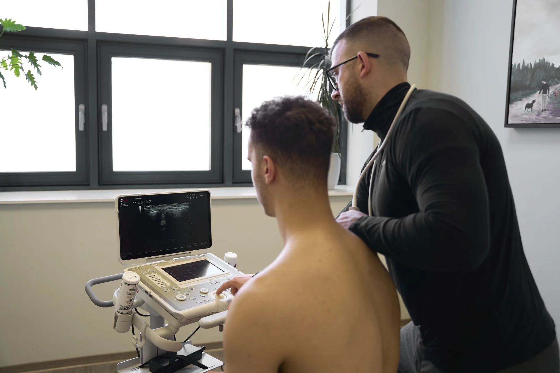
Brief: what do you see with ultrasound?
Ultrasound allows you to image tendons and surrounding structures in detail. Among other things, you can observe the following:
- Thickening or swelling of the tendon
- Tendon degeneration (tendinosis)
- Partial cracks or abnormalities in fibre structure
- Calcifications
- Tendinitis (tenosynovitis)
- Increased blood flow (Doppler) in active symptoms

In what complaints is ultrasound useful?
Ultrasound helps assess:
- Long-term or recurrent tendon complaints
- Suspected partial rupture
- Calcifications or bursa problems in shoulder pain
- Unexplained local pain
- Evaluation of recovery in an existing treatment pathway

Making a difference in treatment
The ultrasound helps determine which approach is appropriate:
- Degenerative tendon without inflammation: exercise therapy aimed at building tendon load (e.g. eccentric exercises)
- Calcifications or bursitis: possibly targeted injection or movement adjustment
- Partial rupture: scheduling rest periods followed by controlled rehabilitation
- Active inflammation (with Doppler activity): anti-inflammatory before build-up
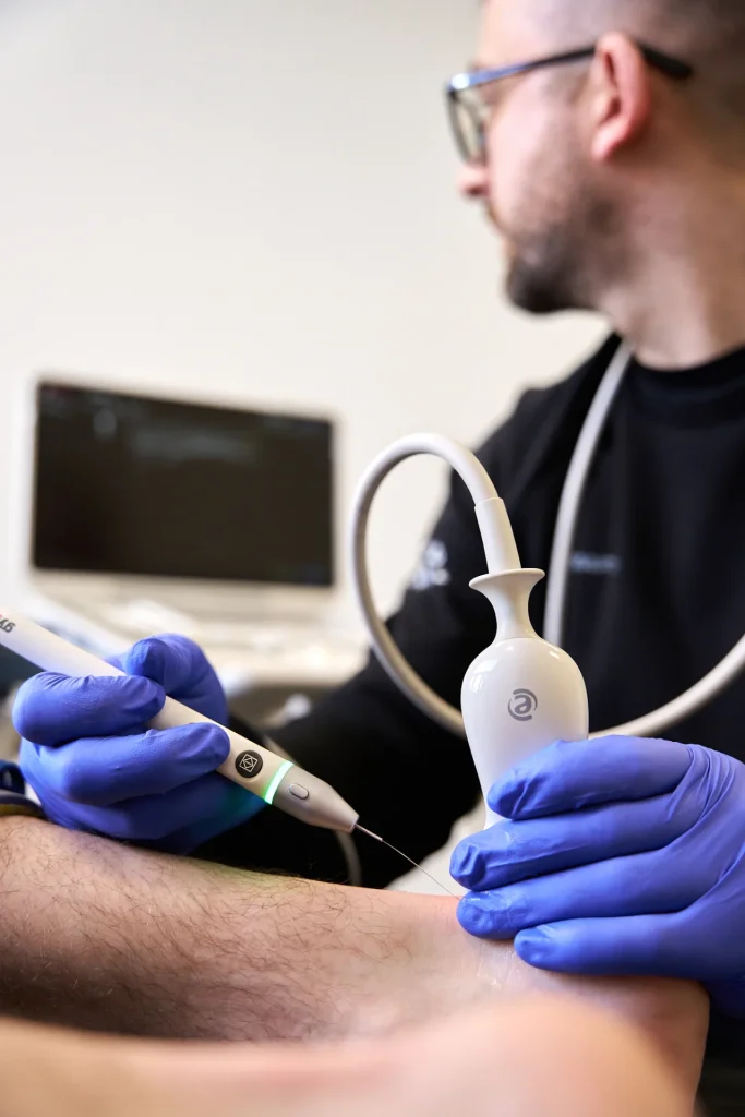
- Chronic tendinopathy with poor recovery:
Then percutaneous electrolysis (PE) can be considered. This involves inserting a thin needle into the affected tendon under ultrasound guidance, after which a weak current activates the local repair process. This technique stimulates inflammation resolution and tendon tissue regeneration, often as an adjunct to exercise therapy.
Case study
An athletic man of 45 comes down with long-term Achilles tendon pain. Rest has helped little so far. Ultrasound shows clear tendon thickening with increased blood flow (Doppler positive) and signs of degeneration. The treatment plan is adjusted: less friction, build up of eccentric load, and additionally several sessions of percutaneous electrolysis. After six weeks, pain is significantly reduced and function increased.
Advice: have an ultrasound if in doubt
Do you have long-term or unclear tendon complaints? Then ultrasound can provide a lot of clarity. It helps to determine what exactly what is going on, and which approach to that.
In short: when in doubt, look with ultrasound. You will see more than you think.
Related complaints

Achilles tendon rupture
A complete rupture of the Achilles tendon often occurs during explosive movements where there is a sudden application of a lot of force,...

Hamstring complaints in runners
Hamstring complaints are common among runners. Pain and loss of strength are common symptoms in hamstring injuries.