Surgery for shoulder instability
The shoulder is a joint that can move in many directions. Because the shoulder joint can move so well, there is a greater risk of the joint dislocating. We call this in the medical world a shoulder luxation. When the shoulder dislocates, there is possible damage to the ligaments, tendons and capsule of the shoulder. This often happens during a quick sudden movement but even more often after a fall. Once dislocated, we see that the shoulder dislocates more easily. The reason for this is that the ligaments and capsule can no longer provide sufficient strength when large forces are applied to the shoulder during sports or other times in daily life. At this point, we speak of shoulder instability. When physiotherapy no longer offers sufficient results, surgery may be necessary to prevent worse.

Possible causes of shoulder luxation
Young athletic people are more likely to suffer shoulder luxation. This is often the result of a fall or collision. When this happens more often, the risk of new injury is higher. Flexibility of the ligaments and tendons can also be congenital. We then speak of hypermobility of the shoulder. Often also genetically determined. People with hypermobility have a higher risk of shoulder dislocation. To some extent, this can be prevented by putting a good load on the muscles and ligaments through strength training. Chronic conditions may also play a role. Ehlers Danlos syndrome increases the risk of shoulder instability because of connective tissue deterioration. Our tendons, ligaments, muscles are all different forms of connective tissue types. In Ehlers Danlos, the connective tissue in our body acquires a high elasticity. Therefore, this significantly increases the risk of luxation.
Diagnosis of shoulder instability
When the shoulder has dislocated more often, the likelihood of persistent shoulder instability is significantly increased. Imaging studies such as an MRI scan, X-ray or CT scan often provide a good picture about the anatomical structures in and around the shoulder.
Some examples of injuries or injuries to the shoulder are:
A bankart lesion. A bankart lesion can occur in two ways: a bony bankart lesion or a normal bankart lesion. A bankart lesion is located in the lower part of the labrum. The labrum is a cartilage-like ring that provides extra stability in the shoulder. This ring, as it were, enlarges the naturally small socket of the shoulder and sits like a suction cup around the head of the shoulder. A bankart lesion is an injury to the labrum. In a bankart lesion, the labrum is damaged or even torn away from the socket of the shoulder. When a piece of bone from the socket tears off along with it, we speak of a bony bankart lesion.
A bankart lesion can occur in several ways. We often see excessive force on the shoulder causing damage to this part of the labrum. A bankart lesion can also be caused by repeated overuse of the shoulder. Think of overhead sports like baseball where there is a lot of force on the shoulder over a long period of time. The pain is usually located at the front of the shoulder and sometimes there is a recognisable click during movements of the shoulder.
Conservative vs surgical
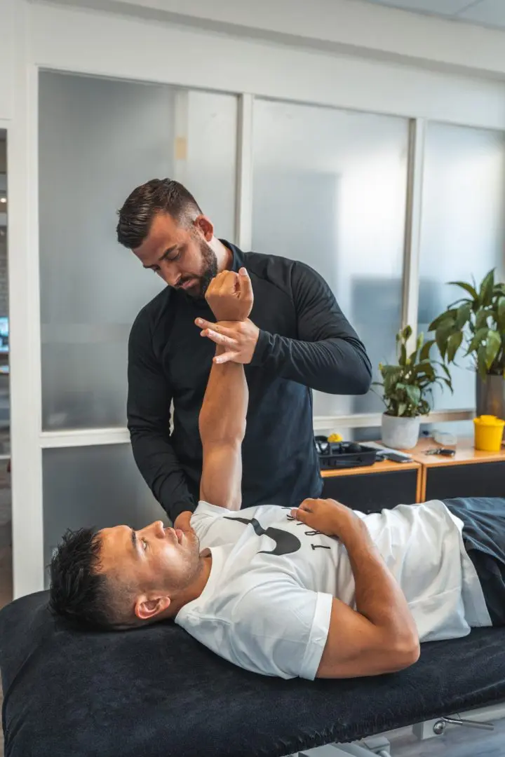
When it is chosen not to operate, physiotherapy is an option. The muscle strength of the shoulder is trained to increase its stability. NSAIDs can also be used to relieve pain. Should the symptoms not diminish sufficiently, surgery may be considered. The damaged lower part of the labrum is reattached. In some cases, the shoulder capsule is also damaged. This will then also be repaired during this operation. After surgery, the shoulder should not be heavily loaded. In the first phase of rehabilitation, the mobility of the shoulder will be restored, before training can resume to increase the load-bearing capacity of the shoulder.
Treatment of a SLAP lesion
Non-operative
A SLAP lesion can be treated with physiotherapy. It then looks at how best to train the shoulder without causing too much discomfort. Pain relief is also often used to suppress symptoms. In some cases, a corticosteroid injection may be chosen. Treatment focuses mainly on increasing muscle strength of the muscles around the shoulder. Overhand activities and overhead sports are discouraged during rehabilitation. In some cases, it is again possible to function pain-free within the chosen sport. If this is not the case after 4-5 months, surgery may still be chosen.
Operative
When physiotherapy does not give the desired result, surgery may be necessary. Through keyhole surgery, the torn part of the biceps attachment is sutured to the socket of the shoulder. Normally, after this procedure, the biceps tendon can resume its function normally. However, research shows that this does not always give the desired effect. A large group of people still have some degree of pain and reduced function of the shoulder even after surgery. A choice can also be made to detach the biceps tendon in its entirety from the place where it attaches to the socket of the shoulder. We call this a biceps tendon tenotomy. This method often gives better results in terms of pain relief.
After surgery
After surgery, a sling (sling) is fitted. This is worn for 4-6 weeks. On average, shoulder rehabilitation after surgery takes 8 months to a year. A person is often unable to drive a car or ride a bike by himself in the first 8 weeks. After 4-6 months, a person can resume participating in various sports. With contact sports, this will take just a little longer.
Physio Fitaal specialises in rehabilitation after surgery. Rehabilitation after surgery often has a big impact and can be exciting. Fysio Fitaal makes rehabilitation after surgery insightful, manageable and transparent. Independence and self-reliance are important pillars within rehabilitation. The team at Fysio Fitaal in Tilburg provides an exclusive service with a personal approach. On your own to a fit and pain-free life. Together with you, we will draw up the rehabilitation plan to help you get closer to your goal.

